CLINICAL EXPERIENCE

Case Studies

Explore clinical cases across our product portfolio, with procedural insights, original imaging and reported outcomes.

Individual clinical experiences are provided for professional information. Reported outcomes do not establish comparative safety or effectiveness. Refer to applicable local authorization and approved Instructions for Use.

Celosia™ · Case report

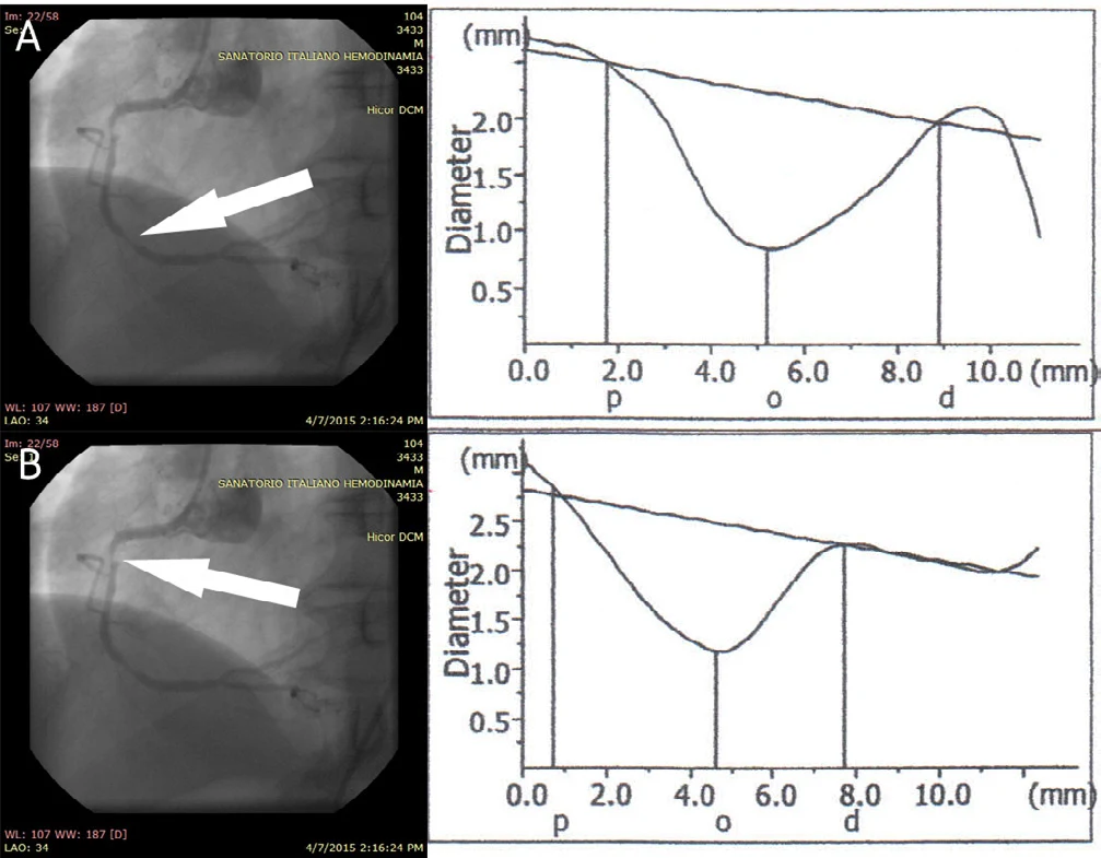
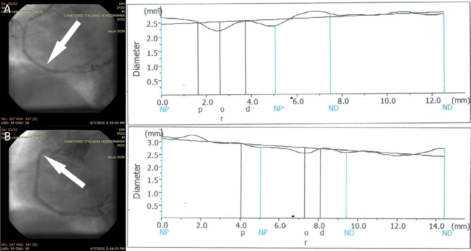
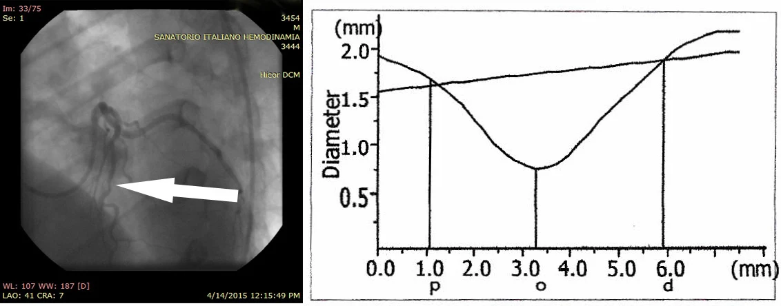
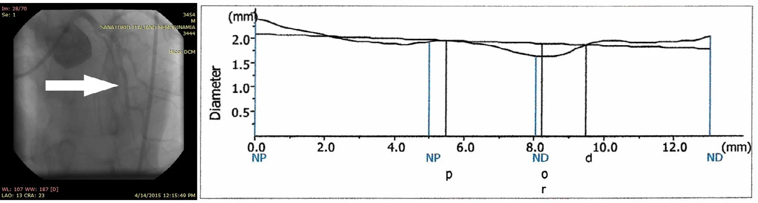
Right coronary artery

Direct stenting of a distal RCA lesion

Presentation

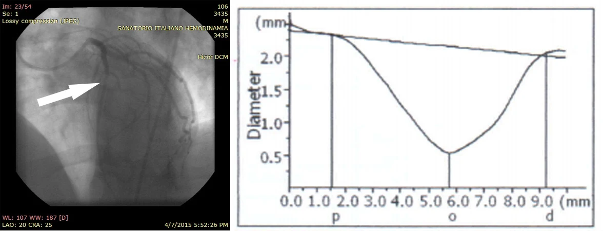
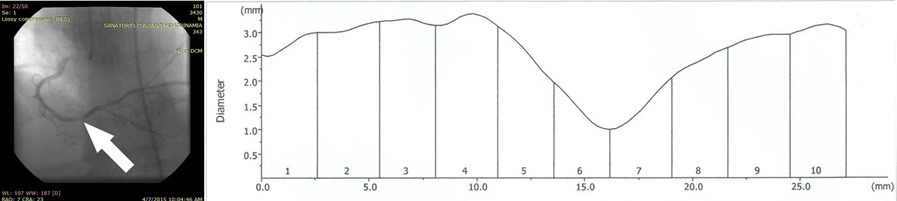
A 55-year-old man with hypertension and a previous myocardial infarction presented with severe distal right coronary artery (RCA) stenosis, described as 88% in the clinical report.

Procedure

A 3.0 × 13 mm Celosia covered stent was placed without balloon pre-dilation, followed by post-dilation.

Reported outcome

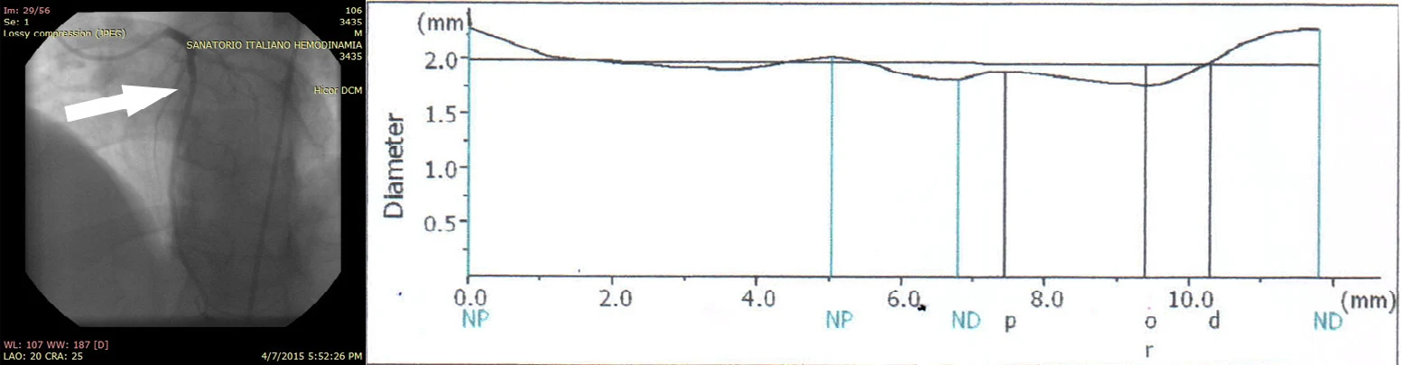
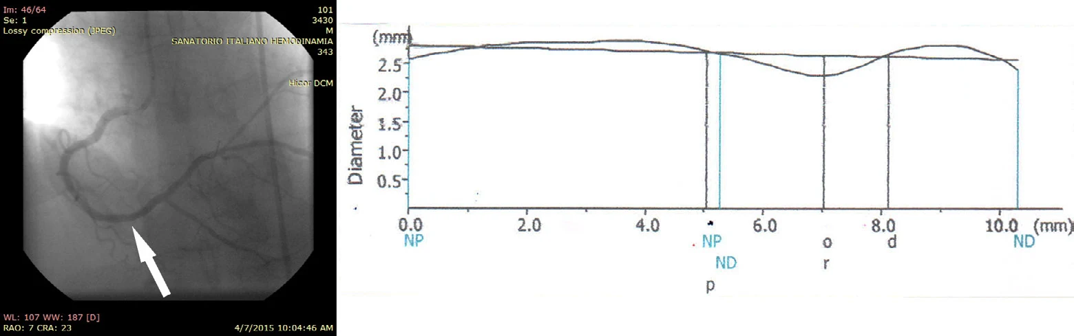
The report describes successful deployment, normal TIMI flow and a post-procedure vessel diameter of 2.57 mm.
Before procedure
Before procedure: angiography and vessel-diameter plot, Right coronary artery
After procedure
After procedure: angiography and vessel-diameter plot, Right coronary artery

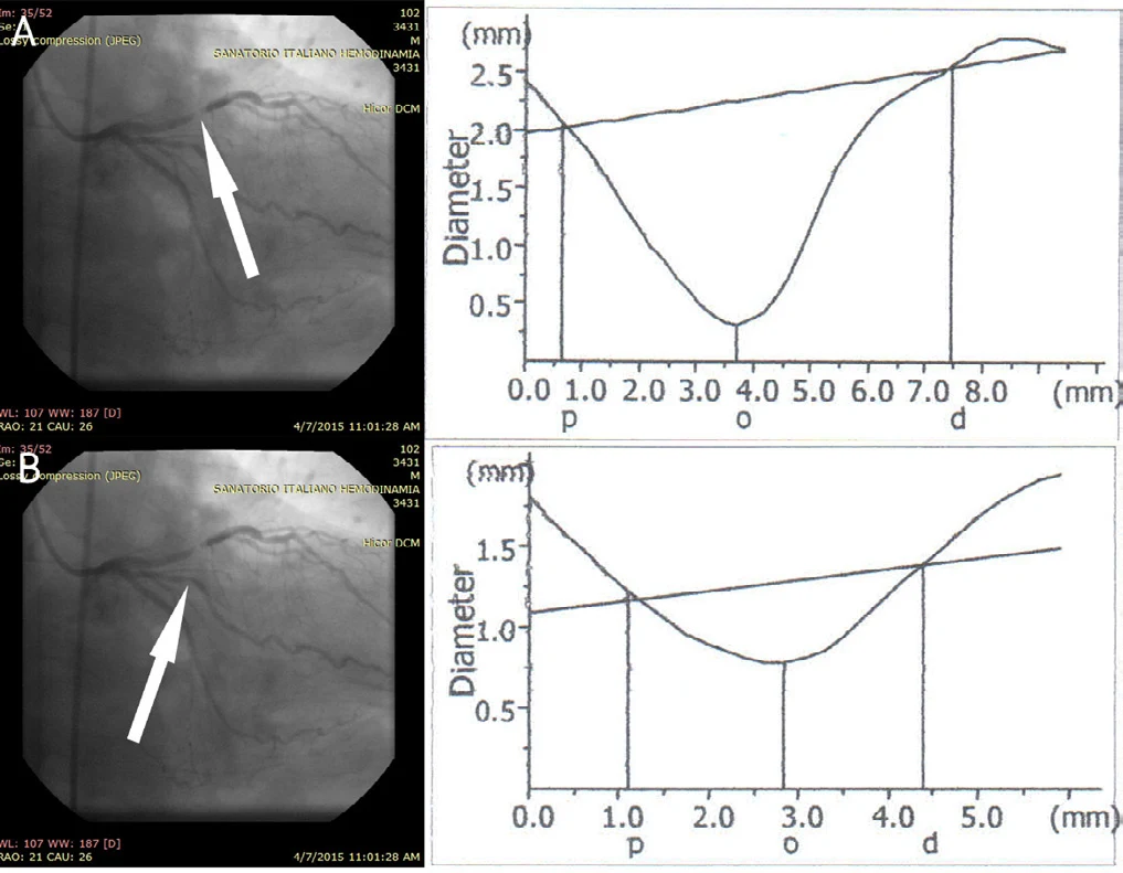
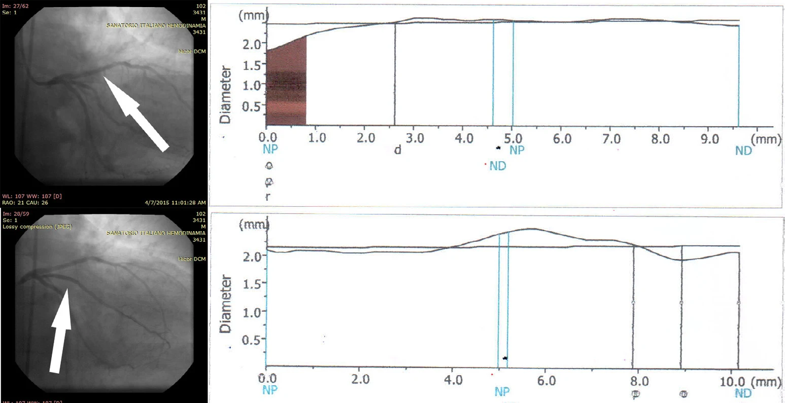
Celosia™ · Case report

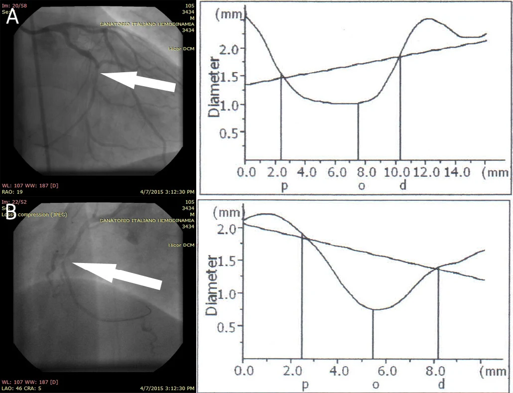
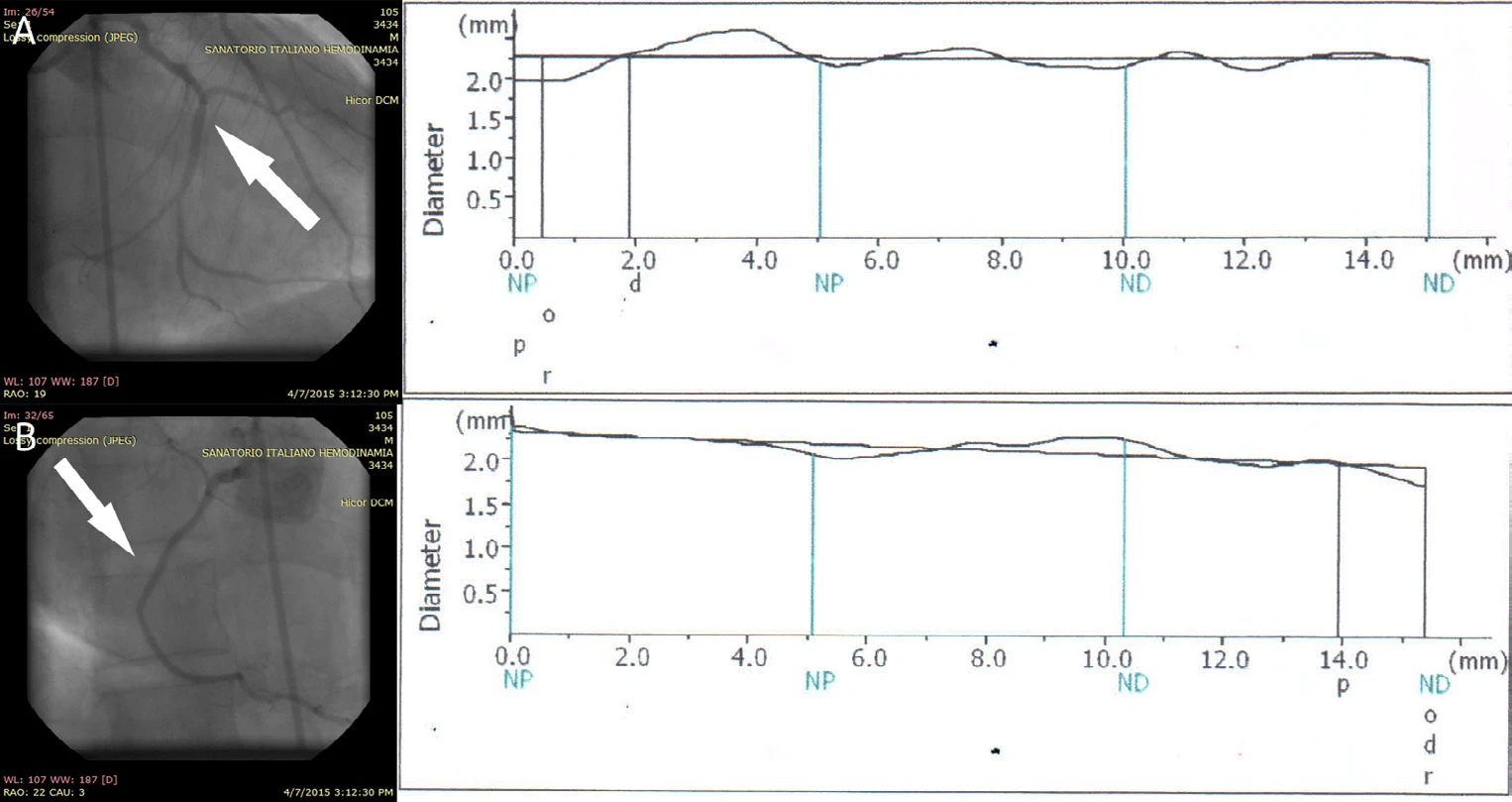
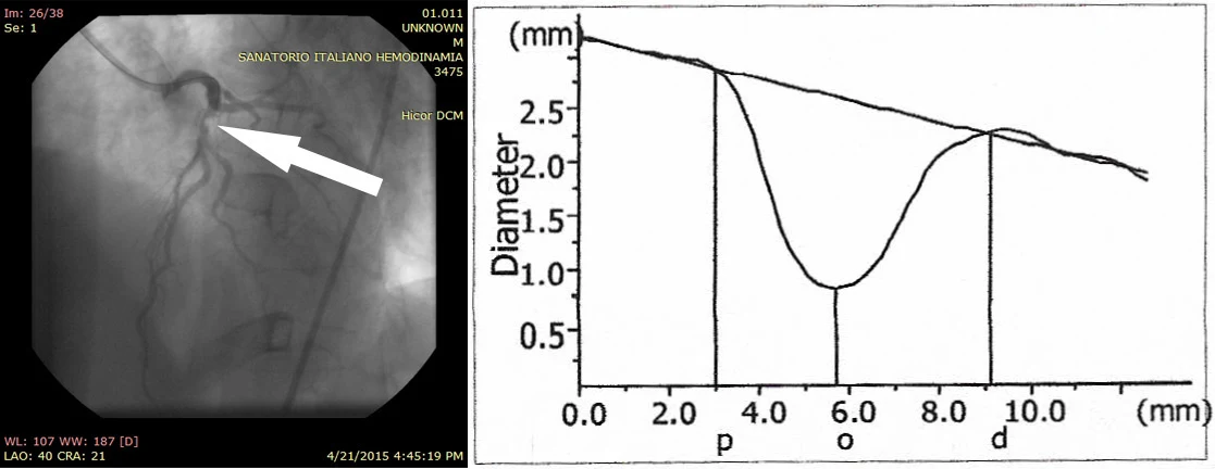
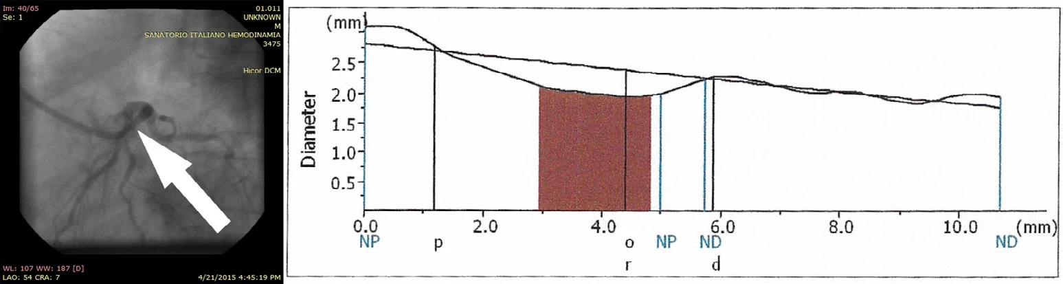
LAD & circumflex branch

LAD treatment and overlapping marginal-branch stents

Presentation

A 61-year-old man with hypertension and diabetes had a 98% proximal left anterior descending (LAD) lesion and a 63% lesion in the first marginal branch of the circumflex artery.

Procedure

The LAD received a 3.0 × 13 mm stent without pre-dilation. The first marginal-branch implant missed part of the lesion, requiring a second covered stent overlapping the first.

Reported outcome

The report describes patent treated vessels, normal TIMI flow and no angiographic migration or dissection after overlapping deployment.

Before procedure
Before procedure: angiography and vessel-diameter plot, LAD & circumflex branch
After procedure
After procedure: angiography and vessel-diameter plot, LAD & circumflex branch

Celosia™ · Case report

Left anterior descending artery

Mid-LAD treatment through tortuous anatomy

Presentation

A 72-year-old woman with hypertension, kyphoscoliosis and spondylosis had a 74% mid-LAD lesion. Tortuosity made passage through the descending aorta challenging.

Procedure

A 2.5 × 16 mm covered stent was advanced to the lesion and deployed without balloon pre-dilation.

Reported outcome

The report describes successful delivery and deployment, normal TIMI flow and a post-procedure mid-LAD diameter of 3.15 mm.

Before procedure
Before procedure: angiography and vessel-diameter plot, Left anterior descending artery
After procedure
After procedure: angiography and vessel-diameter plot, Left anterior descending artery

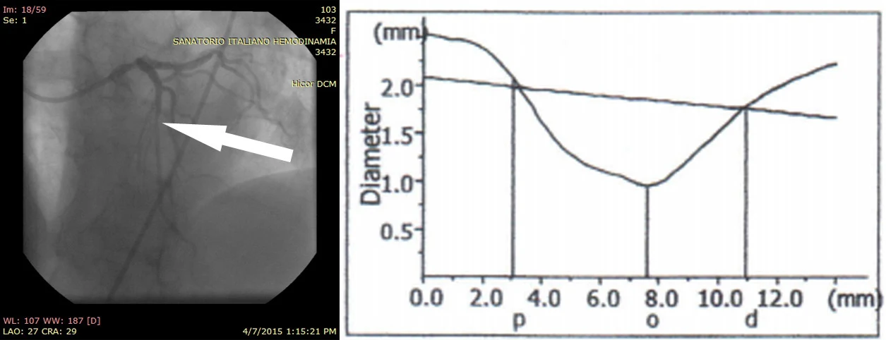
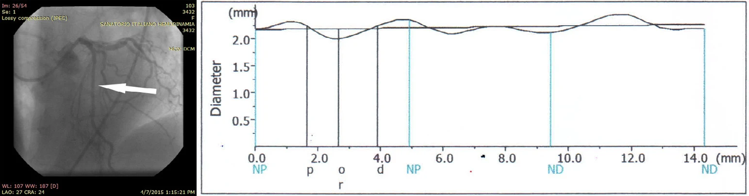
Celosia™ · Case report

Right coronary artery

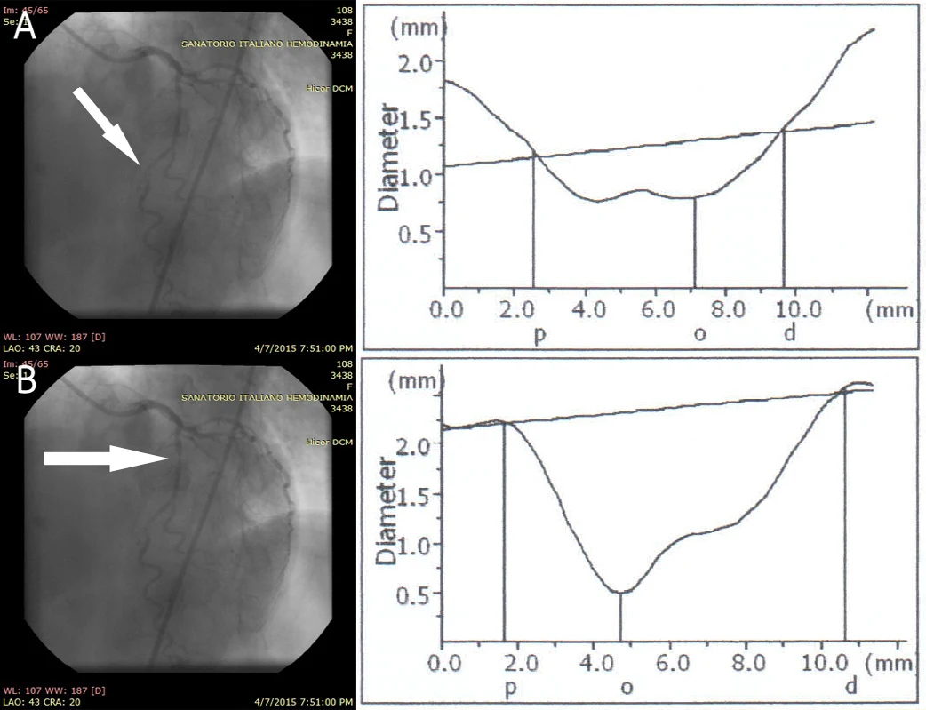
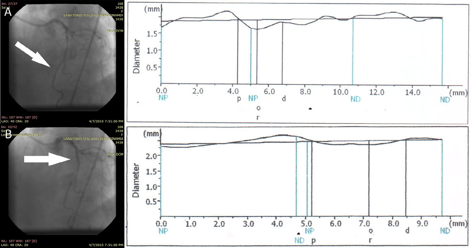
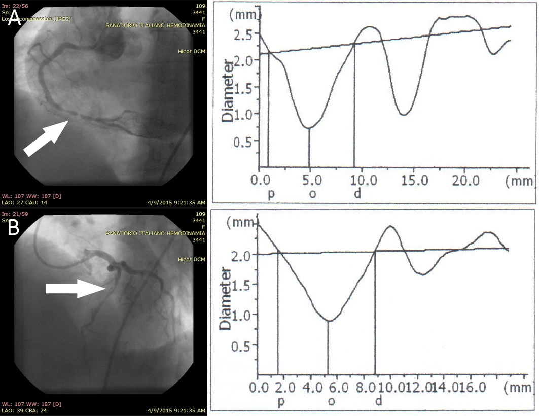
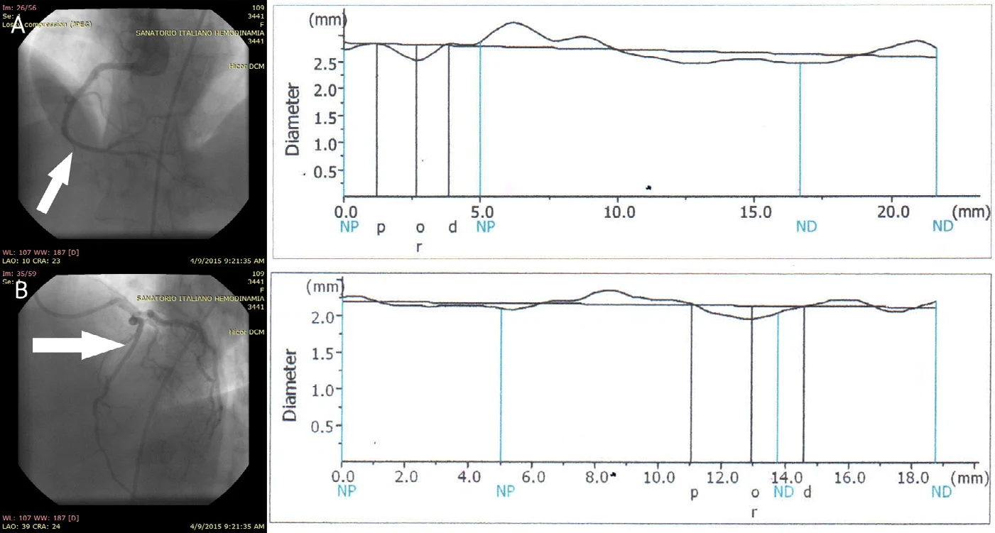
Two RCA lesions treated in one procedure

Presentation

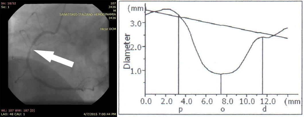
A 51-year-old man with hypertension and previous myocardial infarction had severe thrombotic narrowing in the distal and middle RCA segments.

Procedure

The distal lesion received a 3.0 × 19 mm covered stent, followed by a 3.0 × 13 mm implant in the middle segment. Neither lesion was pre-dilated.

Reported outcome

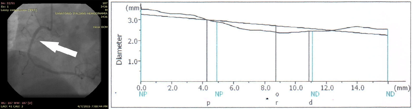
Successful deployment and normal TIMI flow were reported at both sites, with post-procedure diameters of 2.23 mm distally and 2.57 mm in the middle segment.

Before procedure
Before procedure: angiography and vessel-diameter plot, Right coronary artery
After procedure
After procedure: angiography and vessel-diameter plot, Right coronary artery

Celosia™ · Case report

Circumflex & right coronary arteries

Pre-dilation for severe circumflex and RCA disease

Presentation

A 46-year-old man with hypertension and previous myocardial infarction had a 95% circumflex lesion and a 99% RCA lesion with hardened plaque and tortuous anatomy.

Procedure

Both lesions underwent balloon pre-dilation before placement of a 3.0 × 16 mm covered stent in the circumflex and a 3.0 × 19 mm stent in the RCA.

Reported outcome

The report describes successful deployment and normal TIMI flow in both vessels, with post-procedure diameters of 3.05 mm and 2.42 mm respectively.

Before procedure
Before procedure: angiography and vessel-diameter plot, Circumflex & right coronary arteries
After procedure
After procedure: angiography and vessel-diameter plot, Circumflex & right coronary arteries

Celosia™ · Case report

Left anterior descending artery

A proximal LAD lesion requiring pre-dilation

Presentation

A 46-year-old man with hypertension and previous myocardial infarction had a 99% proximal LAD lesion with severe hardened plaque.

Procedure

Initial passage of a 2.5 × 16 mm covered stent was unsuccessful. The device was withdrawn, the vessel was balloon pre-dilated and the stent was reintroduced and deployed.

Reported outcome

The detailed results report successful deployment after pre-dilation, normal TIMI flow and a post-procedure diameter of 1.77 mm.

Before procedure
Before procedure: angiography and vessel-diameter plot, Left anterior descending artery
After procedure
After procedure: angiography and vessel-diameter plot, Left anterior descending artery
Case notes

The report's concluding sentence says no pre-dilation was needed, contradicting its procedure and results. This summary follows the detailed procedural account.

Celosia™ · Case report

Right coronary artery

Direct stenting of a proximal RCA lesion

Presentation

A 56-year-old man with hypertension, diabetes and previous myocardial infarction had a 92% proximal RCA lesion.

Procedure

A 3.0 × 16 mm Celosia covered stent was placed without balloon pre-dilation, followed by post-dilation.

Reported outcome

The report describes successful deployment, normal TIMI flow and a post-procedure proximal RCA diameter of 2.39 mm.

Before procedure
Before procedure: angiography and vessel-diameter plot, Right coronary artery
After procedure
After procedure: angiography and vessel-diameter plot, Right coronary artery

Celosia™ · Case report

Left anterior descending artery

Different preparation strategies for two LAD lesions

Presentation

A 68-year-old woman with hypertension had distal and proximal LAD lesions in tortuous anatomy, described in the diagnostic account as 63% and 96% stenosis.

Procedure

The distal lesion received a 2.5 × 16 mm covered stent directly. Hardened plaque blocked passage at the proximal lesion; balloon pre-dilation preceded placement of a 3.0 × 16 mm stent there.

Reported outcome

The report records normal TIMI flow and post-procedure diameters of 1.64 mm distally and 2.35 mm proximally.

Before procedure
Before procedure: angiography and vessel-diameter plot, Left anterior descending artery
After procedure
After procedure: angiography and vessel-diameter plot, Left anterior descending artery
Case notes

Some source passages label the affected vessel or segment differently. This summary follows the detailed LAD diagnosis and procedure.

Celosia™ · Case report

RCA & left anterior descending artery

Tandem distal RCA lesions and proximal LAD disease

Presentation

A 56-year-old woman with hypertension had two tandem 99% distal RCA lesions and additional proximal LAD disease.

Procedure

The procedure describes balloon pre-dilation in both vessels, followed by a 2.5 × 26 mm covered stent in the distal RCA and a 3.0 × 26 mm stent in the proximal LAD.

Reported outcome

Successful deployment and normal TIMI flow were reported, with post-procedure diameters of 2.53 mm in the RCA and 1.97 mm in the LAD.

Before procedure
Before procedure: angiography and vessel-diameter plot, RCA & left anterior descending artery
After procedure
After procedure: angiography and vessel-diameter plot, RCA & left anterior descending artery
Case notes

The source summary and procedure differ on which lesions were pre-dilated, and LAD severity varies across passages. This summary follows the procedure and omits an ambiguous LAD percentage.

Celosia™ · Case report

Left anterior descending artery

Direct stenting of a mid-LAD lesion

Presentation

A 48-year-old man with hypertension and a family history of coronary artery disease had an 81% mid-LAD lesion, with no previous documented myocardial infarction.

Procedure

A 2.5 × 13 mm covered stent was deployed without balloon pre-dilation, followed by post-dilation.

Reported outcome

The report records successful implantation, normal TIMI flow and a post-procedure vessel diameter of 1.64 mm.

Before procedure
Before procedure: angiography and vessel-diameter plot, Left anterior descending artery
After procedure
After procedure: angiography and vessel-diameter plot, Left anterior descending artery

Celosia™ · Case report

Left anterior descending artery

Direct stenting of a proximal LAD lesion

Presentation

A 69-year-old man with hypertension, a family history of coronary artery disease and previous myocardial infarction presented with a 91% proximal LAD lesion.

Procedure

A 2.5 × 19 mm covered stent was delivered and deployed without balloon pre-dilation, followed by post-dilation.

Reported outcome

The report describes successful implantation, normal TIMI flow and a post-procedure proximal LAD diameter of 1.94 mm.

Before procedure
Before procedure: angiography and vessel-diameter plot, Left anterior descending artery
After procedure
After procedure: angiography and vessel-diameter plot, Left anterior descending artery

Celosia™ · Case report

Left anterior descending artery

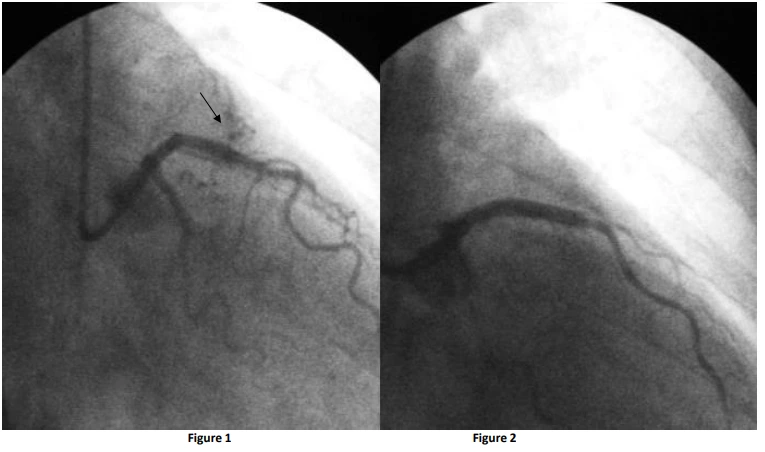
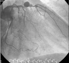
Exclusion of coronary fistulas in the LAD

Presentation

A 62-year-old woman presented with coronary fistulas and complex vessel anatomy. An endovascular approach was selected to exclude the abnormal flow pathways in the left anterior descending (LAD) artery.

Procedure

Under fluoroscopic guidance, a 3.5 × 19 mm covered stent was positioned across the affected LAD segment. The report identifies the implant by Celosia's former name, Direct-Stent Stent Graft.

Reported outcome

Post-deployment imaging showed fistula exclusion, device expansion and stable positioning without immediate complications. At six months, the report describes clinical stability, no recurrence of abnormal flow, sustained vessel patency and maintained device position.

LAD angiography: before (left) and after (right)
Coronary angiography showing LAD fistulas before implantation on the left and their exclusion after implantation on the right

Celosia™ · Case report

Left anterior descending artery

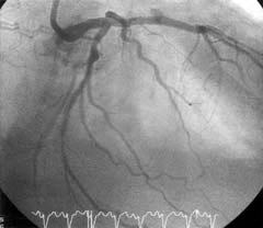
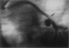
Covered-stent exclusion of a proximal LAD aneurysm

Presentation

An illustrated case in Dr. Bogart Parra's coronary intervention report shows a 10 mm aneurysm in the proximal LAD. Patient demographics and presenting symptoms are not supplied.

Procedure

A 3.0 × 26 mm InSitu Direct-Stent Stent Graft, the former Celosia product name, was implanted across the aneurysm. The report provides paired angiograms documenting the intervention.

Reported outcome

The post-implant angiogram is described as showing sealing of the aneurysm. No patient-specific follow-up interval, longer-term outcome or complication assessment is supplied for this illustrated case.

Before: proximal LAD aneurysm
Figure 3: coronary angiography showing a proximal LAD aneurysm
After: aneurysm exclusion
Figure 4: angiography after a 3.0 × 26 mm InSitu covered stent sealed the aneurysm

Celosia™ · Case report

Saphenous vein graft to the LAD

Treatment of obstruction and aneurysm in a vein graft

Presentation

The illustrated case describes 90% obstruction and an aneurysm in a saphenous vein graft supplying the LAD. The report does not provide patient demographics or presenting symptoms.

Procedure

An InSitu covered stent was implanted in the affected graft. The source uses the former Stent Graft product name; the case is catalogued here under Celosia. Implant dimensions are not supplied for this case.

Reported outcome

A post-implant angiogram is provided alongside the pretreatment image. The figure caption gives no quantitative residual stenosis, follow-up interval or patient-specific clinical outcome.

Before: vein-graft obstruction and aneurysm
Figure 1: saphenous vein graft with 90% obstruction and aneurysm
After covered-stent implantation
Figure 2: angiogram of the vein graft after covered-stent implantation